A new insight into the Brain
New in Dubai: color duplex Sonography of the inner basal brain vessels
The c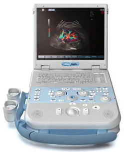 olor duplex Sonography is a very common and often used method in all areas of medicine. In neurology, the method has been used for many years for the study of the carotid arteries that is vessels supplying the brain. It helps evaluate the vessel wall structures and the wall elasticity assess and demonstrate calcifications and deposits (so called plaques) on the vessel walls already at the onset of atherosclerosis. Thus it allows optimal treatment of vascular risk factors at an early stage.
To assess the vascular segments within the brain so far only the CW duplex Sonography was available, which enabled the assessment of stenoses or anomalies on blood flow velocity profiles.
Thanks to recent technological developments, a representation of the inner basal cerebral vessels is now also possible with color duplex Sonography.
By means of a so called pulsed probe that is attached to the temples, through the skull bonesthe basal arteries and veinscan be represented in the image. Together with the measurement of flow velocities (“Duplex”) now also the vessels in the brain can be accurately represented. The investigation is important, for example, for the evaluation of headache, especially migraine (e.g., vascular malformations), dizziness, after – or better – before – strokes.
Furthermore, also some areas of the brain can be represented, so that e.g.Parkinson’s disease can be detected at an early stage or various forms of Parkinson’s disease can be distinguished from each other – what makes a targeted therapy possibleright from the start.
The examination is painless, takes about 10 to 15 minutes and is now available in the German Neuroscience Center.